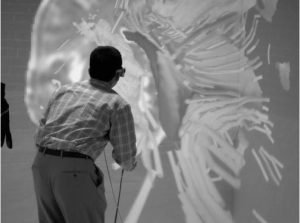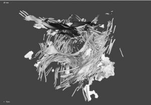


Studying changes in white-matter and pre-operative planning for brain tumor surgery are the two applications supported by this system. Usually such analysis is done through 2D slice reconstructed from the DT-MRI data but the use of a CAVE allows to render a 3D model of the brain. This model consists in streamtubes which are thin tubes that extend from seed points following the direction of fastest diffusion.
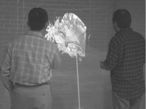
Moreover, the use of CAVE is claimed to allow collaboration between expert around the same 3D model. Communication is managed naturally since both users are in the same space and can see each other entirely, however for pin-point gesture a raycasting solution has been employed since only one user have the correct distortion making hand-pointing difficult.